




| IN A NUTSHELL |
|
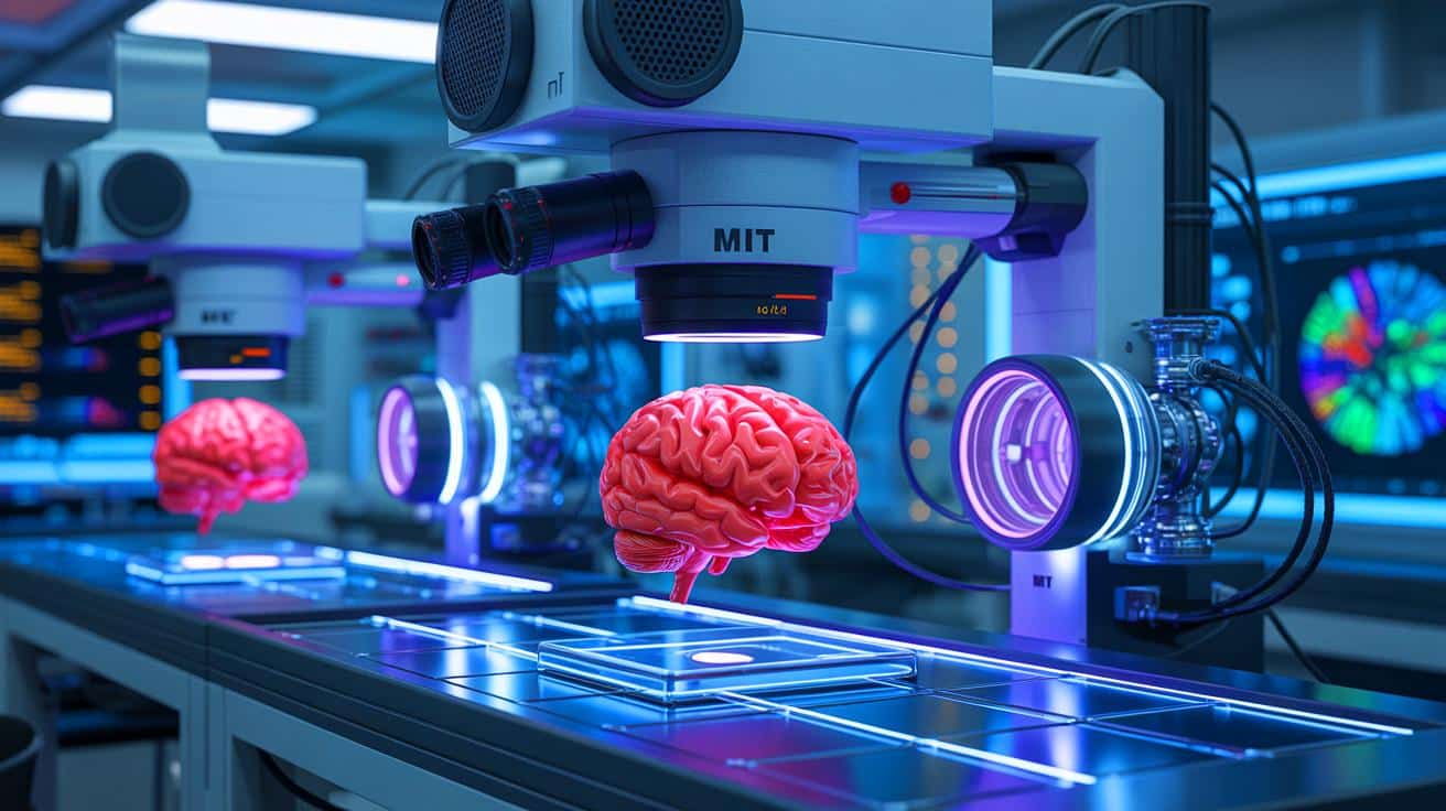
In a groundbreaking development, researchers at MIT have unveiled a novel imaging system capable of penetrating brain tissue five times deeper than previous technologies. By utilizing a unique combination of light and sound waves, this innovative system provides unprecedented insights into the brain’s complex structure without altering the cells. This breakthrough holds the potential to revolutionize neuroscience research and surgical applications, offering new avenues for understanding brain function and diseases. As scientists continue to push the boundaries of microscopy, this latest advancement marks a significant step forward in the quest to map the brain’s intricate networks more accurately and comprehensively.
Seeing Deeper Into the Brain
The newly developed imaging system at MIT represents a significant leap forward in the field of neuroscience. The system is capable of detecting NAD(P)H, a crucial molecule linked to cell metabolism and neuronal activity, through dense brain samples. This was demonstrated in tests involving a 1.1-millimeter thick human stem cell-derived cerebral organoid and a 0.7-millimeter slice of mouse brain tissue. The samples were not large enough to fully test the system’s limits, according to W. David Lee, a postdoctoral researcher involved in designing the system.
The core technology of this system lies in its use of a three-photon excitation method. This method fires ultrashort light bursts at triple the molecule’s normal absorption wavelength, allowing the light to scatter less and penetrate deeper into the tissue. The absorbed energy causes a rapid, microscopic thermal expansion inside the cell, which generates sound waves. These waves are then detected by a sensitive ultrasound microphone, and software algorithms convert them into high-resolution images. This process, known as three-photon photoacoustic imaging, enables researchers to visualize deeper brain structures without the need for dyes or genetic modification.
Merging Advanced Imaging Techniques
The MIT team has successfully integrated three-photon excitation, photoacoustic detection, and label-free imaging into a single platform dubbed “Multiphoton-In and Acoustic-Out.” This system allows for precise molecular detection without altering the tissue. The platform can also identify other molecules, such as GCaMP, a calcium indicator used to track neural activity. Furthermore, the system employs “third-harmonic generation” imaging to map cellular structures, providing both structural and molecular details in a single scan.
Co-lead author Tatsuya Osaki from The Picower Institute emphasized the goal of combining advanced techniques into one efficient process. This innovation could prove instrumental in studying conditions where NAD(P)H levels fluctuate, including Alzheimer’s disease, Rett syndrome, and seizures. Additionally, because the system operates without labels, it holds the potential to guide brain surgeries by mapping activity in real time. The next steps involve testing the system in living animals, where both the light source and microphone will need to be positioned on the same side of the tissue.
Potential Applications and Future Directions
The implications of this new imaging technology extend beyond basic research. Lee anticipates that the system could image up to 2 millimeters deep in live brains. “In principle it should work,” he noted. The potential applications of this technology are vast, ranging from guiding neurosurgical procedures to advancing the understanding of brain disorders. Lee’s previous work through Precision Healing Inc. demonstrated NAD(P)H imaging’s utility in guiding wound treatment, and the same principles may prove valuable in brain research and neurosurgery.
This project has received funding from several prestigious sources, including the National Institutes of Health, the Simon Center for the Social Brain, and The Picower Institute. As the system undergoes further testing and refinement, it is poised to become a critical tool in the arsenal of neuroscientists and surgeons alike. The ability to visualize brain activity at such depths without invasive techniques could transform how we approach neurological diseases and their treatment.
Challenges and Considerations
Despite its promising potential, the new imaging system also presents certain challenges and considerations. Implementing this technology in clinical settings will require addressing technical hurdles, such as ensuring the system’s compatibility with existing surgical tools and protocols. Additionally, the cost and accessibility of the technology will play crucial roles in its widespread adoption.
Researchers will need to conduct extensive testing to validate the system’s efficacy and safety in various contexts. The transition from laboratory to clinic is often fraught with obstacles, necessitating collaboration between scientists, engineers, and medical professionals. However, if successfully implemented, this imaging system could redefine standard practices in both research and clinical environments, offering new hope for patients with neurological conditions.
As this pioneering imaging system continues to evolve, it raises an important question: How will these advances in brain imaging technology impact the diagnosis and treatment of neurological disorders in the coming years?